BMRB Entry 15644
Click here to enlarge.
PDB ID:
Entry in NMR Restraints Grid
Validation report in NRG-CING
Chem Shift validation: AVS_anomalous, AVS_full, LACS
BMRB Entry DOI: doi:10.13018/BMR15644
MolProbity Validation Chart
NMR-STAR file interactive viewer.
NMR-STAR v3 text file.
NMR-STAR v2.1 text file (deprecated)
XML gzip file.
RDF gzip file.
All files associated with the entry
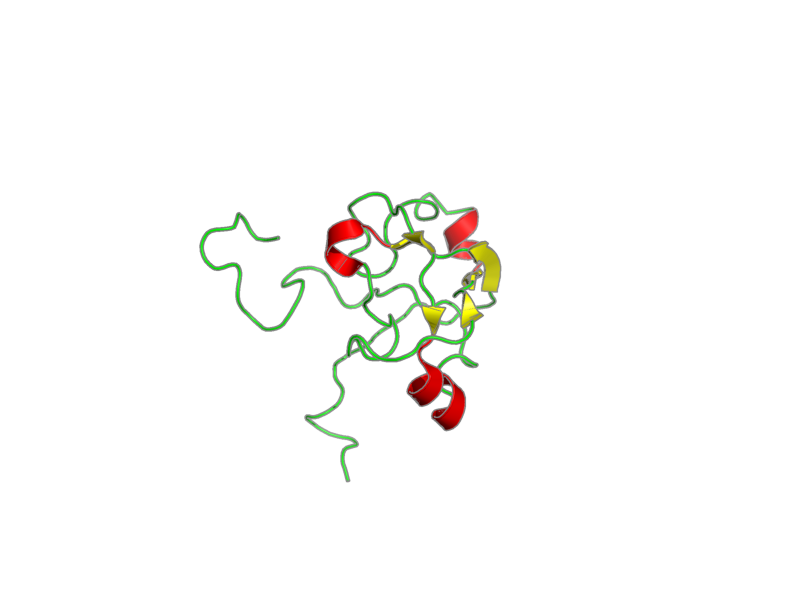
Title: 1H, 15N and 13C chemical shift assignments for Rds3 protein PubMed: 18621724
Deposition date: 2008-01-31 Original release date: 2008-07-28
Authors: Loening, Nikolaus; van Roon, Anne-Marie; Yang, Ji-Chun; Nagai, Kiyoshi; Neuhaus, David
Citation: van Roon, Anne-Marie; Loening, Nikolaus; Obayashi, Eiji; Yang, Ji-Chun; Newman, Andrew; Hernandez, Helena; Nagai, Kiyoshi; Neuhaus, David. "Solution structure of the U2 snRNP protein Rds3p reveals a knotted zinc-finger motif" Proc. Natl. Acad. Sci. U. S. A. 105, 9621-9626 (2008).
Assembly members:
Rds3p, polymer, 109 residues, 12353.605 Da.
ZN, non-polymer, 65.409 Da.
Natural source: Common Name: baker Taxonomy ID: 4932 Superkingdom: not available Kingdom: not available Genus/species: Eukaryota Fungi
Experimental source: Production method: recombinant technology Host organism: Escherichia coli
Entity Sequences (FASTA):
Rds3p: GGSSRHQFDLIMCLKQPGVQ
TGLLCEKCDGKCPICDSYVR
PKRKVRVCENCSFGKQAKNC
IICNLNVGVNDAFYCWECCR
LGKDKDGCPRILNLGSNRLD
RHFEKKKKV
- assigned_chemical_shifts
| Data type | Count |
| 13C chemical shifts | 479 |
| 15N chemical shifts | 109 |
| 1H chemical shifts | 756 |
Additional metadata:
Assembly:
| Entity Assembly ID | Entity Name | Entity ID |
|---|---|---|
| 1 | Rds3 | 1 |
| 2 | ZINC ION_1 | 2 |
| 3 | ZINC ION_2 | 2 |
| 4 | ZINC ION_3 | 2 |
Entities:
Entity 1, Rds3 109 residues - 12353.605 Da.
residues -1 to 1 (GGS) result from cleavage of an N-terminal GST fusion protein
| 1 | GLY | GLY | SER | SER | ARG | HIS | GLN | PHE | ASP | LEU | ||||
| 2 | ILE | MET | CYS | LEU | LYS | GLN | PRO | GLY | VAL | GLN | ||||
| 3 | THR | GLY | LEU | LEU | CYS | GLU | LYS | CYS | ASP | GLY | ||||
| 4 | LYS | CYS | PRO | ILE | CYS | ASP | SER | TYR | VAL | ARG | ||||
| 5 | PRO | LYS | ARG | LYS | VAL | ARG | VAL | CYS | GLU | ASN | ||||
| 6 | CYS | SER | PHE | GLY | LYS | GLN | ALA | LYS | ASN | CYS | ||||
| 7 | ILE | ILE | CYS | ASN | LEU | ASN | VAL | GLY | VAL | ASN | ||||
| 8 | ASP | ALA | PHE | TYR | CYS | TRP | GLU | CYS | CYS | ARG | ||||
| 9 | LEU | GLY | LYS | ASP | LYS | ASP | GLY | CYS | PRO | ARG | ||||
| 10 | ILE | LEU | ASN | LEU | GLY | SER | ASN | ARG | LEU | ASP | ||||
| 11 | ARG | HIS | PHE | GLU | LYS | LYS | LYS | LYS | VAL |
Entity 2, ZINC ION_1 - Zn - 65.409 Da.
| 1 | ZN |
Samples:
sample_1: Rds3p, [U-98% 15N], 350 uM; Tris buffer, [U-99% 2H], 20 mM; sodium chloride 200 mM; DTT, [U-99% 2H], 1 mM
sample_2: Rds3p, [U-98% 13C; U-98% 15N], 700 uM; Tris buffer, [U-99% 2H], 20 mM; sodium chloride 200 mM; DTT, [U-99% 2H], 1 mM
sample_conditions_1: ionic strength: 200 mM; pH: 7.0; pressure: 1 atm; temperature: 300 K
Experiments:
| Name | Sample | Sample state | Sample conditions |
|---|---|---|---|
| 2D 1H-15N HSQC | sample_1 | isotropic | sample_conditions_1 |
| 2D 15N T1 | sample_1 | isotropic | sample_conditions_1 |
| 2D 15N T2 | sample_1 | isotropic | sample_conditions_1 |
| 2D 15N NOE | sample_1 | isotropic | sample_conditions_1 |
| 2D 1H-15N HSQC | sample_2 | isotropic | sample_conditions_1 |
| 2D 1H-13C HSQC full width | sample_2 | isotropic | sample_conditions_1 |
| 2D 1H-13C HSQC aromatic | sample_2 | isotropic | sample_conditions_1 |
| 2D 1H-13C HSQC aliphatic | sample_2 | isotropic | sample_conditions_1 |
| 2D 1H-1H NOESY | sample_2 | isotropic | sample_conditions_1 |
| 2D 1H-1H NOESY filtered (15N coupled 1H removed from F2) | sample_2 | isotropic | sample_conditions_1 |
| 3D HNCACB | sample_2 | isotropic | sample_conditions_1 |
| 3D CBCA(CO)NH | sample_2 | isotropic | sample_conditions_1 |
| 3D HNCO | sample_2 | isotropic | sample_conditions_1 |
| 3D HNHAHB | sample_2 | isotropic | sample_conditions_1 |
| 3D HBHA(CO)NH | sample_2 | isotropic | sample_conditions_1 |
| 3D H[C]CH-TOCSY | sample_2 | isotropic | sample_conditions_1 |
| 3D [H]CCH-TOCSY | sample_2 | isotropic | sample_conditions_1 |
| 3D HNHB | sample_1 | isotropic | sample_conditions_1 |
| 3D 1H-15N NOESY | sample_2 | isotropic | sample_conditions_1 |
| 3D 1H-13C NOESY | sample_2 | isotropic | sample_conditions_1 |
| 3D HACAHB-COSY | sample_2 | isotropic | sample_conditions_1 |
Software:
xwinnmr v3.5, Bruker Biospin - processing
CCPN_analysis v1.0.15, CCPN - data analysis
ARIA v1.1.2, Linge, O, . - refinement
CNS v1.1, Brunger, Adams, Clore, Gros, Nilges and Read - structure solution
NMR spectrometers:
- Bruker Avance 800 MHz
- Bruker DMX 600 MHz
- Bruker DRX 500 MHz
Related Database Links:
| PDB | |
| DBJ | GAA27055 |
| EMBL | CAY87048 |
| GB | AAB68140 AAT93199 AHY78252 AJP42220 AJV91308 |
| REF | NP_015419 |
| SP | Q06835 |
| TPG | DAA11509 |
Download simulated HSQC data in one of the following formats:
CSV: Backbone
or all simulated shifts
SPARKY: Backbone
or all simulated shifts