BMRB Entry 17689
Click here to enlarge.
PDB ID:
Entry in NMR Restraints Grid
Validation report in NRG-CING
Chem Shift validation: AVS_full, SPARTA
BMRB Entry DOI: doi:10.13018/BMR17689
MolProbity Validation Chart
NMR-STAR file interactive viewer.
NMR-STAR v3 text file.
NMR-STAR v2.1 text file (deprecated)
XML gzip file.
RDF gzip file.
All files associated with the entry
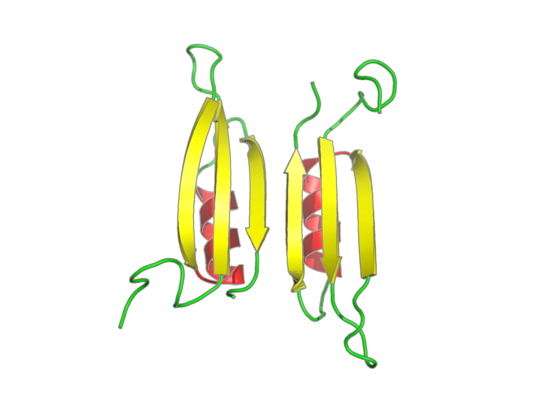
Title: Novel dimeric structure of phage phi29-encoded protein p56: Insights into Uracil-DNA glycosylase inhibition PubMed: 21890898
Deposition date: 2011-06-06 Original release date: 2012-02-22
Authors: Asensio, Juan Luis; Perez-Lago, Laura; M. Lazaro, Jose; Gonzalez, Carlos; Serrano-Heras, Gemma; Salas, Margarita
Citation: Asensio, Juan Luis; Perez-Lago, Laura; Lazaro, Jose; Gonzalez, Carlos; Serrano-Heras, Gemma; Salas, Margarita. "Novel dimeric structure of phage 29-encoded protein p56: insights into uracil-DNA glycosylase inhibition." Nucleic Acids Res. 39, 9779-9788 (2011).
Assembly members:
p56, polymer, 56 residues, 6571.369 Da.
Natural source: Common Name: Bacteriophage phi-29 Taxonomy ID: 10756 Superkingdom: Viruses Kingdom: not available Genus/species: Bacteriophage phi29
Experimental source: Production method: recombinant technology Host organism: Escherichia coli
Entity Sequences (FASTA):
p56: MVQNDFVDSYDVTMLLQDDD
GKQYYEYHKGLSLSDFEVLY
GNTADEIIKLRLDKVL
- assigned_chemical_shifts
| Data type | Count |
| 15N chemical shifts | 58 |
| 1H chemical shifts | 391 |
Additional metadata:
Assembly:
| Entity Assembly ID | Entity Name | Entity ID |
|---|---|---|
| 1 | p56 chain A | 1 |
| 2 | p56 chain B | 1 |
Entities:
Entity 1, p56 chain A 56 residues - 6571.369 Da.
| 1 | MET | VAL | GLN | ASN | ASP | PHE | VAL | ASP | SER | TYR | ||||
| 2 | ASP | VAL | THR | MET | LEU | LEU | GLN | ASP | ASP | ASP | ||||
| 3 | GLY | LYS | GLN | TYR | TYR | GLU | TYR | HIS | LYS | GLY | ||||
| 4 | LEU | SER | LEU | SER | ASP | PHE | GLU | VAL | LEU | TYR | ||||
| 5 | GLY | ASN | THR | ALA | ASP | GLU | ILE | ILE | LYS | LEU | ||||
| 6 | ARG | LEU | ASP | LYS | VAL | LEU |
Samples:
sample_1: p56 0.5 mM; sodium chloride 100 mM; potassium phosphate 10 mM; H2O 90%; D2O 90%
sample_2: p56, [U-98% 13C; U-98% 15N], 0.5 mM; sodium chloride 100 mM; potassium phosphate 10 mM; H2O 90%; D2O 90%
sample_3: p56 0.5 mM; sodium chloride 100 mM; potassium phosphate 10 mM; H2O 90%; D2O 90%
sample_conditions_1: ionic strength: 0.13 M; pH: 5; pressure: 1 atm; temperature: 308 K
Experiments:
| Name | Sample | Sample state | Sample conditions |
|---|---|---|---|
| 2D 1H-1H TOCSY | sample_1 | isotropic | sample_conditions_1 |
| 2D 1H-1H NOESY | sample_1 | isotropic | sample_conditions_1 |
| 2D 1H-1H COSY | sample_1 | isotropic | sample_conditions_1 |
| 2D 1H-1H NOESY | sample_3 | isotropic | sample_conditions_1 |
| 2D 1H-15N HSQC | sample_2 | isotropic | sample_conditions_1 |
| 3D HNCACB | sample_2 | isotropic | sample_conditions_1 |
| 3D CBCA(CO)NH | sample_2 | isotropic | sample_conditions_1 |
Software:
XEASY, Bartels et al. - data analysis
DYANA, Guntert, Braun and Wuthrich - structure solution
AMBER, Case, Darden, Cheatham, III, Simmerling, Wang, Duke, Luo, ... and Kollm - refinement
NMR spectrometers:
- Bruker Avance 800 MHz
- Bruker DMX 600 MHz
Download simulated HSQC data in one of the following formats:
CSV: Backbone
or all simulated shifts
SPARKY: Backbone
or all simulated shifts