|
Biological Magnetic Resonance Data BankA Repository for Data from NMR Spectroscopy on Proteins, Peptides, Nucleic Acids, and other Biomolecules |
Member of
|
Ub Mediated Proteolysis |
|||||
| |||||
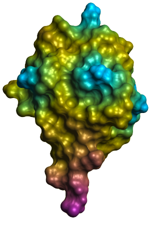
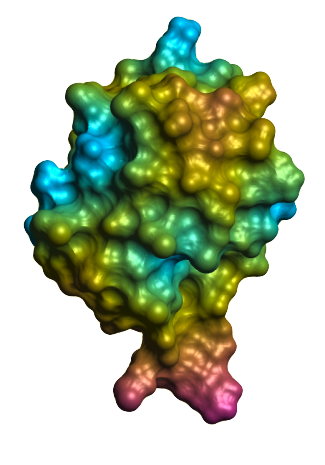
The best understood and possibly most important function of Ub is to tag specific proteins for deconstruction by the 26S proteasome. The 26S proteasome is a large, multimeric barrel shaped protein with regulatory caps on either end. Alhough the interior of the barrel is highly proteolytic, the exterior is benign to its surroundings. The caps can recognize a ubiquinated protein, de-ubiquinate and denature it, and then allow it to pass through the core. Left alone, proteins tend to degrade over time. Ub-mediated proteolysis requires energy in the form of ATP. It might seem odd that a cell would devote energy to destroying a molecule that it just invested energy in building. However, most of the proteins targeted in this way are molecules that are needed for short periods of time, and that would be deleterious to the cell if they lingered too long. They are often regulatory proteins themselves that need to be quickly turned on and off. Sometimes the targeted proteins are simply mistakes, which have been either mistranslated or misfolded. As one would expect, Ub is ligated to a target protein by a specific reaction. The first step is ATP-dependent and involves the formation of a thiolester bond between the cysteine of a Ub-activating enzyme, known as an E1 enzyme, and the Ub C-terminus. The E1 enzyme can then transfer the ubiquitin to a Ub-conjugating enzyme (E2). The E2 enzyme works in conjunction with a Ub-ligating enzyme (E3) to form a covalent amide linkage to a lysine on the target protein. It is the E2/E3/Ub/target interaction that specifies which proteins are degraded and the rates at which they are degraded. Although Ub is highly conserved, the E2 and E3 enzymes are highly variable.
Once the targeted protein has been ubiquitinated, it isn't necessarily tagged for destruction. Its fate is usually specified by whether or not the one or more Ub molecules are attached to the first to form a poly-ubiquin chain. Ub has seven lysine residues, to which the C-terminal of another Ub can be attached. The most important, as far as proteolysis is concerned is residue Lys(48), though Lys(11) and Lys(29) poly-ubiquinated chains also tag a protein for proteolysis. Poly-ubiquitination is also mediated by E3 ligases: some consider these particular ligases as a separate class of Ub-elongation enzymes (E4). It is really quite an elegant system. The E2 and E3 enzymes make up a library of protein regulators. They can label the protein as ready for recycling by a generic proteasome, or they can tag the protein for some other purpose. They themselves do not need any mechanism to interact with the proteasome or anything other than Ub and their target substrates. The European Bioinformatics Institue on their December 2004 Protein of the Month provides a good overview of some of the other roles ubiquitin plays within the cell. | |||||
|
Previous: Introduction |

